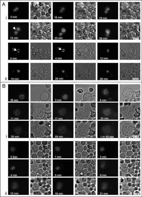
Figure 2.
Chromosomal segregation patterns differ in high and low ploidy MKs. Several movies were recorded with imaging intervals of 90–121 seconds (see Suppl. Movies under Suppl. Material). (A) A low ploidy MK undergoing endomitosis. (A, part i) MK chromosomal condensation is evident during and up to eighteen minutes, with DNA assuming a tight, compact and lobulated form. At nineteen minutes chromosomes formed two groups that moved towards the poles of the cell, and at twenty-five minutes (not shown here, but followed in the movies) they rejoined into a single cluster. The shape of the MK also changed from spherical to elongated and reverted back to spherical at the last steps of chromosomal re-gathering into one nucleus (for measurements of cell size and the distance between chromosomes see Suppl. Figs. 1 and 2). (A, part ii) A low ploidy MK captured at anaphase and undergoing final steps of an endomitotic cycle. Initially, the two chromosomal groups were separated by space consistent with midzone formation (indicated by white arrows) and gradually re-gathered into a single group (within about fourteen minutes). (B, part i and ii) High ploidy MKs (16N, 8N) captured at early mitosis. Chromosomal condensation was followed by transient grouping of chromosomes at 8–9 minutes, followed by spreading into a ring type alignment with formation of territories between the chromosomes, reaching maximum size at about fifteen minutes after condensation (for measurements of cell size and the distance between chromosomes see Suppl. Figs. 3–7). The fluorescent intensity seems somewhat reduced at latter stages of anaphase, as expected when the chromosomes (and fluorescence) are more spread (compare 36 sec to 1 h 50 min in B, part i). Some cell furrowing is observed in the 8N cell (see phase microscope movie SM4A at 16 min), but typically not in higher ploidy cells. Data shown represent analysis of eight time-lapse microscopy sessions, each focused on a single MK.

