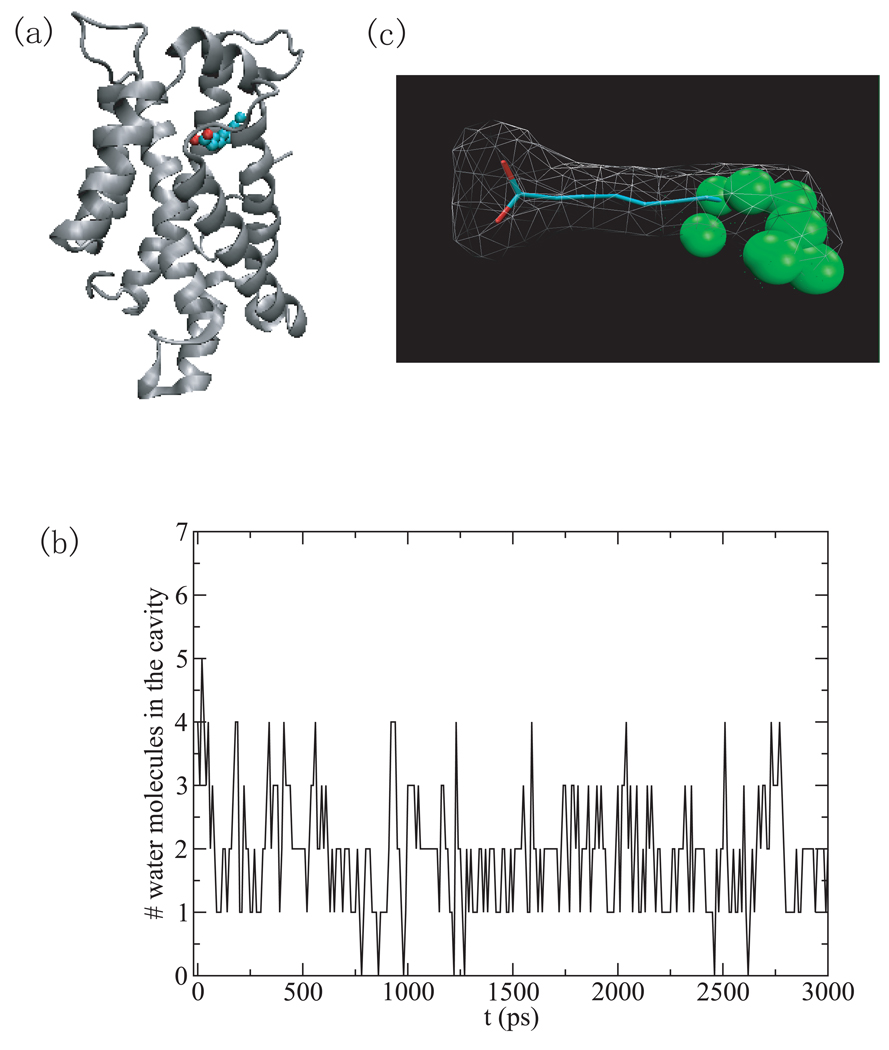
Fig.3.
(a) The structure of the bovine apo-glycolipid transfer protein (PDB ID 1wbe) is shown as cartoon and the bound fatty acid, decanoic acid, is shown in ball and stick representation. (b) The number of water molecules within 2Å of the decanoic acid versus simulation time in the hydrophobic channel. (c) Water density in the hydrophobic channel.

