
Figure 13.
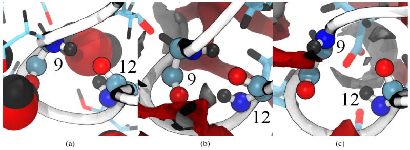
Backbone hydrogen bonding between residues 9 and 12 for representative structures of (a) explicit SPC/E, (b) 3D-RISM-KH with hydrogen bonding and (c) 3D-RISM-KH without hydrogen bonding. Protein backbone drawn as a white tube, backbone atoms for residues 9 and 12 as spheres and side chains as sticks. Carbons are cyan, oxygens red, hydrogens black and nitrogens blue. Solvent density isosurfaces are shown at for both oxygen (red) and hydrogen (gray). Images made with VMD.71,72