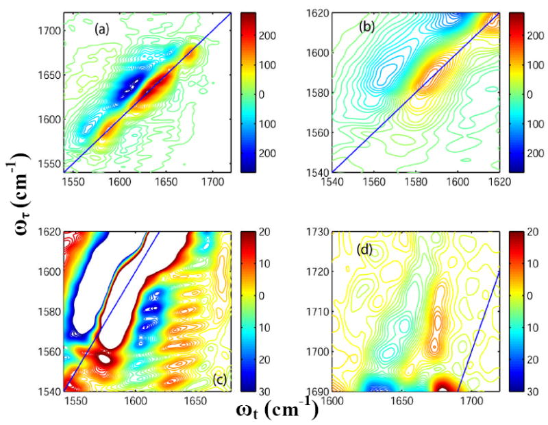
Figure 4. 2D-IR spectra of Villin headpiece at 23° C.

Real part of the absorptive 2D-IR spectrum of HP35 in neat D2O (T = 0) at 23° C focusing on: (a) the whole frequency range for the carbonyl stretches; (b) the diagonal peaks from Asp and Glu ionizable side chains; (c) the cross peaks arising due to the coupling between the ionizable side chains and the main amide-I bands; and (d) the cross peaks arising between the COOH side chain of Asp and Glu and the TFA.
