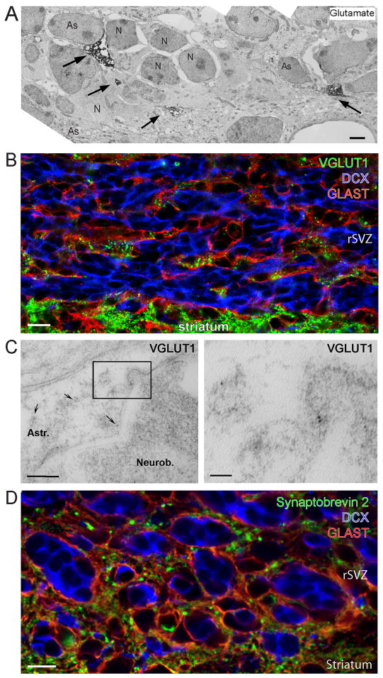
Figure 3. VGLUT1 and Synaptobrevin 2 are only expressed in astrocytes in the SVZ/RMS.
(A) Electron micrograph of pre-embedding immunocytochemistry for L-glutamate in the rSVZ. The electron dense material for glutamate (as indicated by the arrows) is observed exclusively in astrocytes (As) surrounding neuroblasts (N). Scale: 1 μm. (B) Confocal image displaying co-immunostaining for VGLUT1 (green), doublecortin (DCX, blue), and GLAST (red) in the RMS. Scale: 10 μm. (C) Electron micrographs showing post-embedding immunocytochemistry for VGLUT1 expression in the rSVZ. The right panel represents a higher magnification image of the boxed region shown in the left panel. The secondary antibody was conjugated to 5 nm-gold particles (arrows). Scale bars: 200 nm (left)/50 nm (right). (D) Confocal image of synaptobrevin 2 (green), DCX (blue), and GLAST (red). Scale: 10 μm.

