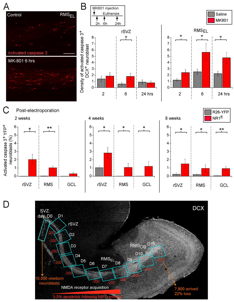
Figure 7. In vivo blockade and genetic removal of NMDARs decrease neuroblast survival.
(A) Confocal images displaying immunostaining for activated caspase 3 (red) in the RMS from control or MK-801-treated DCX-GFP mice. Scale: 100 μm. (B) Bar graphs of the density of activated caspase 3-immunopositive DCX+ cells in sagittal slices containing the rSVZ and RMSEL following in vivo injections of saline solution (Ctl) or MK-801. Mice received a single dose of MK-801 and were sacrificed at different time-points following injection (2, 6, and 24 hrs). The density was obtained by dividing the cell number by the volume of the region analyzed. The inset illustrates the experimental paradigm. (C) Bar graphs of the percentage of activated caspase 3+ YFP+/DCX+ neuroblasts in the rSVZ, RMS and granule cell layer (GCL) of the OB quantified at 2, 4, and 8 weeks post-electroporation. The % was calculated from the total number of YFP+ neuroblasts. (D) Model illustrating the predicted effect of increased apoptosis on accumulated cell loss along the migratory route. Considering that ~10,000 neuroblasts are born per day, ~200 NR1-KO neuroblasts (1–3%) will be apoptotic and cleared in ~1 day. Since its takes ~11 days for newly born neuroblasts to migrate from the SVZ to the RMSOB, apoptosis would lead to a total loss of ~2,200 neuroblasts in the RMSOB, corresponding to 22% of the original neuroblast population.

