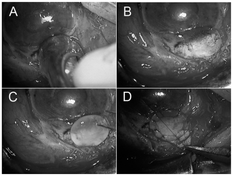
Figure 2.
Using a corneal trephine (5.5 mm diameter), half-layered corneoscleral tissues was dissected away (A). Clean, healthy edges of the dissection were regularized (B). Then a donor’s lamellar, round, preserved corneal piece with 5.5 mm diameter and 0.8 mm thickness was placed (C) and was sutured exactly to the patient’s corneoscleral area with 10-0 nylon (D).

