Fig. 3.
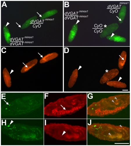
A dVGAT mutant. Embryos were derived from dVGATminos1/CyO parents, and visualized using a standard upright (A–D) or confocal (E–J) microscope. Embryos from two separate low-power fields are shown in A,C and B,D. For all panels, anti-dVGAT, labeling is in red and endogenous fluorescence of GFP is green. Intense GFP fluorescence (A,B white arrowheads) is seen in embryos homozygous for the GFP-tagged minos insertion (dVGATminos1/dVGATminos1). These embryos do not show specific labeling for dVGAT in the ventral nerve cord (C,D, white arrowheads). Embryos showing less intense GFP fluorescence (A,B, white arrows) are heterozygous for the mutation (dVGATminos1/CyO) and show robust labeling for dVGAT in the nerve cord (C,D, white arrows). The remaining, malformed embryo shown in B and D (asterisk) is presumably CyO/CyO. (E–G) Confocal images of additional embryos. A dVGATminos1/CyO heterozygote (E–G) shows moderate GFP fluorescence (E, white arrow) and labeling of dVGAT in the ventral nerve cord (F, white arrow). A dVGATminos1/dVGATminos1 homozygote shows high levels of GFP fluorescence (H, arrowhead) and no specific labeling of dVGAT in the nerve cord (I, white arrowhead). As seen in the merged images (G,J), both heterozygotes and homozygotes show non-specific autofluorescence in the gut (small grey arrowheads). Scale bars, 100 μm.

