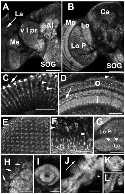
Fig. 5.
Expression of dVGAT in the adult optic ganglia and brain. (A,B) Frontal views of an adult fly brain and optic lobes. An anterior optical section (A) shows punctate labeling in the glomeruli of the antennal lobe (Al), the subesophageal ganglion (SOG), ventral lateral protocerebrum (v l pr), and in the optic lobe, the lamina (La) and medulla (Me). A more posterior optical section (B) shows labeling of the lobula (Lo) and lobula plate (Lo P). Higher magnification images show morphologically distinct centrifugal projections of the C2 (C, arrow) and C3 cells (C, arrowheads) in the distal lamina. Labeling in the outer (D, o) and inner medulla (D, i) represent centrifugal processes from C2 and C3 cells, centripetal, putative transmedullary cells (D, arrowhead) (Sinakevitch and Strausfeld, 2004), as well as tangential GABAergic processes (Meyer et al., 1986; Sinakevitch and Strausfeld, 2004) (D, arrow). The retinotopic orientation of labeled processes in the outer medulla can be seen in a view toward the surface of the medulla (E), and is in part derived from the multiple, putative Tm cells (F) whose cell bodies are in the cortical rind of the medulla. (G) Thin processes (arrowheads) may traverse the space between the lobula plate (Lo P), labeled in a loose meshwork pattern, and the lobula (Lo) which appears more stratified than the lobula plate in this view. (H–L) Higher power images of the central brain show labeling of antennal lobe (H) and ellipsoid body (I). Labeled figures likely to be cell bodies are observed adjacent to the antennal lobe (H, arrows). (J) A stack of confocal images shows the dorsal aspect of the thoracic ganglion (arrow points anteriorly). Immunoreactive processes appear to be contained in at least one set of nerve roots (asterisk). Additional views show the prothoracic (K, ventral view) and abdominal (L, ventral view) segments of the ganglion. Scale bars, (A,B,G,J,K,L) 50 μm; (C,D,E,F,H,I) 20 μm.

