
Figure 12.
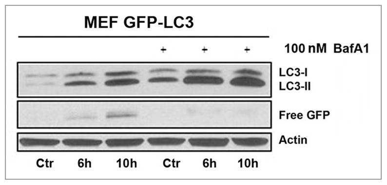
Flux analysis in MEFs stably expressing GFP-LC3. Whole cell lysates were made at the indicated time points and analyzed by western blots. The increased amount of LC3-II and the absence of ‘free GFP’ when cells were pre-treated with 100 nM Bafilomycin A1 1 h before PDT (with hypericin as the photosensitizer) indicates the autophagic flux. All controls represent cells incubated with hypericin in the dark.