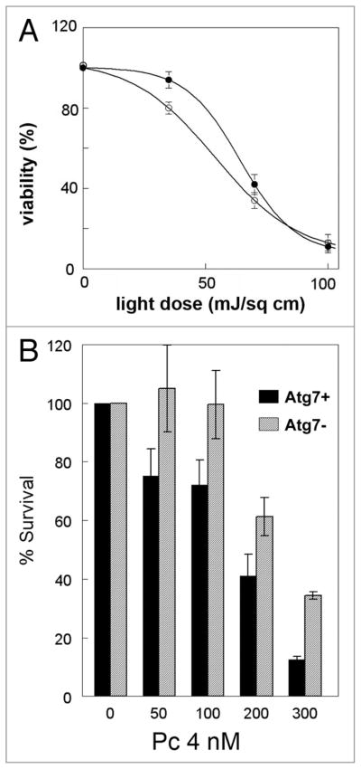
Figure 7.
(A) PDT efficacy in wild-type(●) L1210 cells and an Atg7 knockdown derivative of L1210 cells (○). Cultures of L1210 cells or a L1210 derivative whose expression of Atg7 had been stably knocked down with shRNA were sensitized with CPO and subsequently exposed to light. After photoirradiation cells were plated for assessment of colony formation. Data represent means ± SD of three plates per treatment group. (B) PDT efficacy in wild-type MCF-7 cells (black bars) and in cells whose expression of Atg7 has been stably knocked down (gray bars). Exponentially growing cultures of each cell line were exposed to the indicated concentrations of Pc 4 in complete growth medium for 18 h followed by 200 mJ/cm2 red light. Immediately after photoirradiation, the cells from each dish were collected by trypsinization, and aliquots were replated in 60-mm dishes in sufficient number to result in 50–100 colonies per dish. After 10–14 days, colonies were stained with 1% crystal violet, and colonies having at least 50 cells were counted by eye and normalized to the number of cells plated and the plating efficiency of the control untreated cells. Data for the two cell lines were compared by Student’s t test and found to be significantly different at all doses (p < 0.01).

