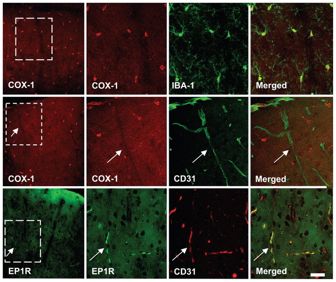
Figure 6.
In somatosensory cortex, COX-1 immunoreactivity (red) is co-localized with the microglial marker Iba-1 (green)(top row), but not with the endothelial marker CD31 (green) (middle row, arrows). EP1 receptor (EP1R) immunoreactivity (green) is co-localized with CD31 (red) (lower row, arrows). The cortical surface is at the top of the panels. The second, third and fourth panels in each row represent enlargements of the square in the first panel. Calibration bar: 10μm.

