
Figure 2.
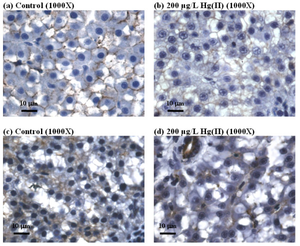
Comparison of hepatic histopathological changes in cell-cell adhesion in control liver (a, c) and induced by HgCl2 (b and d). (a) and (b) show immunohistochemical staining for E. cadherin (indicated by dark brown precipitates). (c) and (d) show immunohistochemical staining for cytokeratin (indicated by dark brown precipitates).