Figure 1.
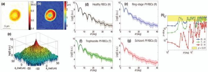
(a) Amplitude and (b) phase map of a healthy RBC. (c) The retrieved light scattering pattern of the same cell. Light-intensity scattering patterns of (d) healthy RBCs, (e) ring, (f) trophozoite, and (g) schizont stage of Pf-RBCs. (h) p-values of scattering patterns (different intraerythrocytic stages of Pf-RBCs are compared to the healthy RBCs).

