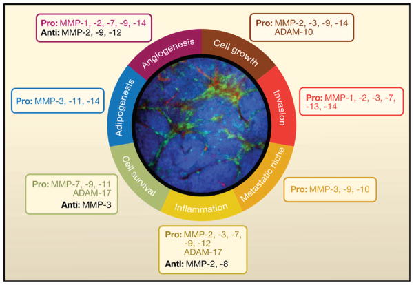
Figure 4. Modulation of the Tumor Microenvironment by MMPs.
Summary of the various processes that are modulated by MMPs in the tumor microenvironment. The selected examples of MMPs and ADAMs promote (pro) or suppress (anti) these processes. An intravital microscopy image of the mammary gland of MMTV-PyMT mice that spontaneously develop mammary carcinoma was taken using a spinning disc inverted confocal microscope (Egeblad et al., 2008). These mice also express enhanced cyan fluorescent protein (CFP) under the control of the actin promoter (ACTB-ECFP) to enable tumor cell labeling (blue) and enhanced green fluorescent protein (GFP) expression under the control of a c-fms promoter (c-fms-EGFP) to label myeloid cells (green). These mice were injected intravenously with 70 kDa rhodamine-dextran to visualize blood vessels (red). This image illustrates the complexity of the tumor microenvironment, which is largely influenced by nonmalignant cells, such as myeloid cells, all of which could be targets as well as sources for MMPs.

