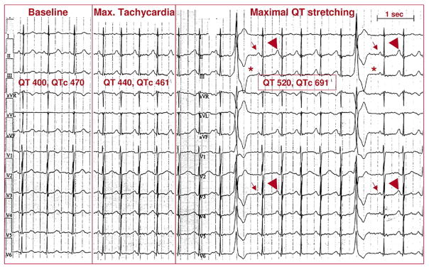
Figure 4. Provocation of Ventricular Arrhythmias by Standing in LQTS.
A 34-year-old woman with previous cardiac arrest and documented pause-dependent torsades de pointes. (Left panel) At baseline, heart rate is 83 beats/min, QT interval is 400 ms, and corrected QT interval (QTc) is 470 ms. (Middle panel) During maximal tachycardia (8 s after standing), heart rate is 115 beats/min, QT interval is 440, and QTc interval is 461 ms. (Right panel) Ventricular extrasystoles appear during maximal QT interval stretching 15 s after standing. Note that the post-extrasystolic pauses (*) expose the small amplitude of P waves (arrows). It is therefore evident that during sinus tachycardia there is a late component of a very long QT interval on top of the P-wave (arrowheads).

