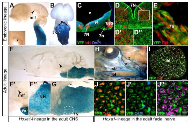
Fig. 3. Hoxa1-lineage contributes extensively to the facial, abducens and mlf nuclei and the facial nerve.
(A–E) Hoxa1-lineage in the embryonic nervous system. (A) Lateral view of a Hoxa1-IRES-Cre; R26R-lacZ E12.5 brain, with Hoxa1-lineage in the caudal brainstem and the medial longitudinal fasciculus (mlf) at the fore/midbrain boundary (arrowhead). Inset is a higher magnification of staining in the nucleus and tracts (arrow) of the mlf. (B) Ventral view of a brain from an E14.5 Hoxa1-IRES-Cre; nLacZ embryo, showing Hoxa1-lineage in the ventral brainstem including the facial nucleus (7N), as well as in migrating pontine nuclei (Pn). (C) Transverse section through the hindbrain of an E11.5 embryo at the level of rhombomere 4 (r4) showing the facial nucleus (7N) and nerve (7n) and the facio-acoustic ganglion complex (7/8G) (lineage in green, DAPI in blue, Isll in red). (D–D″) Higher magnifications of an adjacent section showing strong contribution of Hoxa1-lineage to neurons of the facial nucleus co-stained with Isll (red). (E) Higher magnification of Hoxa1-lineage in axons of the facial nerve co-stained with Tuj1 (red). (F–J″) Hoxa1-lineage in the adult nervous system. (F) Hoxa1-lineage in a sagittal section through the adult brain. (F′, F″) Higher magnification of X-gal positive cells in the mlf and the abducens nucleus (6N). (G) Hoxa1-lineage in the adult seventh nucleus. (H–J″) Hoxa1-lineage in the facial nerve. (H) X-gal staining of an adult Hoxa1-IRES-Cre; R26R head, showing the lineage in all branches of the facial nerve. (I) Cross-section through the inferior buccolabial branch (ibl) of the seventh nerve (dotted line in H) (Hoxa1-lineage in green Tuj1 in red). Higher magnification shows colocalization of Hoxa1-lineage with the neuronal marker Tuj1 (red) (J) and DAPI (blue), which marks Schwann cell nuclei (J′). Myelin basic protein (MBP) immunostaining (magenta) highlights the myelin sheet of Schwann cells (J″). Abbreviations: mm, marginal mandibular; sbl, superior buccolabial; t, temporal; v, ventricle; z, zygomatic. Scale bars in B, F, H: 500 μm; scale bars in C, I: 100 μm.

