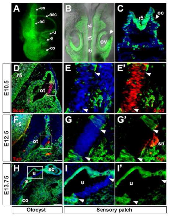
Fig. 5. Hoxa1-lineage contributes extensively to the inner ear but is excluded from sensory regions.
Lineage analysis of Hoxa1-IRES-Cre; R26R-EYFP embryos. (A–C) Hoxa1-lineage (green) in the inner ear, otic vesicle and otic cup. (A) Dissected inner ear of an E13.5 embryo showing strong contribution of Hoxa1-lineage. (B) Dorsal view of an E9.5 embryo with Hoxa1-lineage in the otic vesicle (ov) but excluded from an anterodorsolateral patch (arrowhead). (C) The lineage-label can be detected as early as the otic cup (oc) stage at E8.75 (DAPI, blue). (D–I′) Strong Hoxa1-lineage contribution to the developing otocyst (ot) in transverse sections of E10.5, E12.5 and E13.75 embryos. Hoxa1-lineage is absent from an anterodorsolateral region that was identified as a sensory patch by Sox2 expression (red) which marks sensory cells in the otic epithelium (D) and Tuj1 (red) which marks sensory neurons (sn) and axons which innervate this patch (F). In more differentiated inner ears at E13.75 Hoxa1-lineage is absent from the utricular (u) sensory patch (H). (E, G, I) Higher magnification of the boxed areas in D, F, H, with DAPI counterstain (blue) (E, G, I) or YEP only (E′, G′, I′), showing absence of the lineage from the sensory patches. Arrowheads indicate the region that is devoid of Hoxa1-lineage. Abbreviations: asc, anterior semicircular canal; co, cochlea; ed, endolymphatic duct; es, endolymphatic sac; r4-r6, rhombomeres 4–6; s, saccule; sc, semicircular canals. All scale bars are 100 μm except A: 1mm and B: 200 μm.

