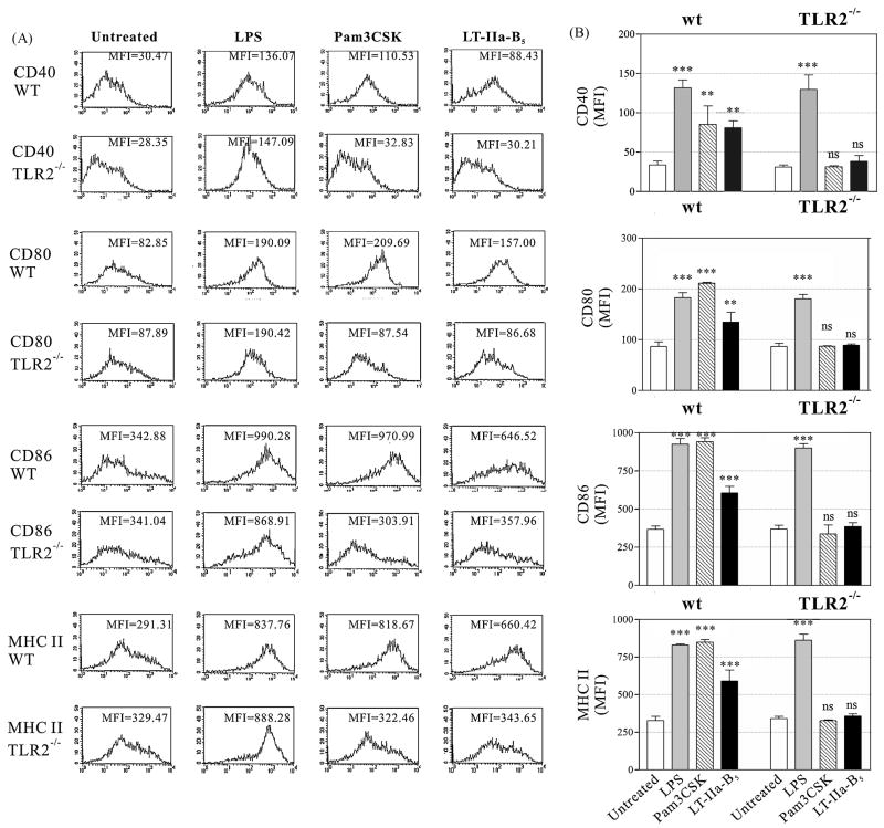
Figure 4. Expression of surface co-stimulatory receptors by wt and TLR2-deficient BMDC after treatment with LT-IIa-B5.
BMDC (1×10⁶) derived from wt C57BL/6 mice or C57Bl/6(TLR2-/-) mice were incubated for 24 hr at 37°C with, PBS (Untreated), 1.0 μg/ml of LPS, 1.0 μg/ml Pam3CSK, or 5.0 μg/ml of LT-IIa-B5. CD11c+ cells were analyzed by flow cytometry for expression of CD40, CD80, CD86, and MHC-II. A. Histographic analysis of expression of CD40, CD80, CD86, and MHC-II. Data from one of three independent experiments are shown. The MFI values for CD40, CD80, CD86, and MHC-II are denoted within each histogram. B. Graphical comparison of the expression of CD40, CD80, CD86, and MHC-II. Error bars denote one standard deviation from the mean. Key: wt, BMDC from TLR2-proficient C57Bl/6 mice; TLR2-/-, BMDC from TLR2-deficient mice; ***, statistical difference from the untreated control (P<0.001); **, statistical difference from the untreated control (P<0.01); ns, not significant.

