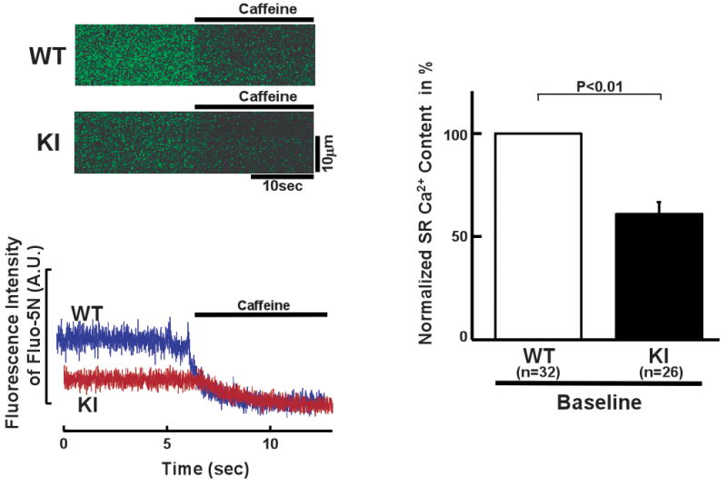
Figure 6.

Spectroscopic determination of luminal [Ca2+] of the SR in saponin-permeabilized WT and R2474S/+ KI cardiomyocytes (see “Expanded Materials and Methods” in online data supplements for measurement of intra-SR [Ca2+]). (Left) Representative fluo-5N images before and after addition of caffeine (20 mmol/L). (Right) Summarized data of the luminal [Ca2+] normalized to control condition of WT cardiomyocytes. N: the number of cells from 3–4 hearts.
