
Figure 2. HGF-induced motility and migration in MDA-MB-231 cells.
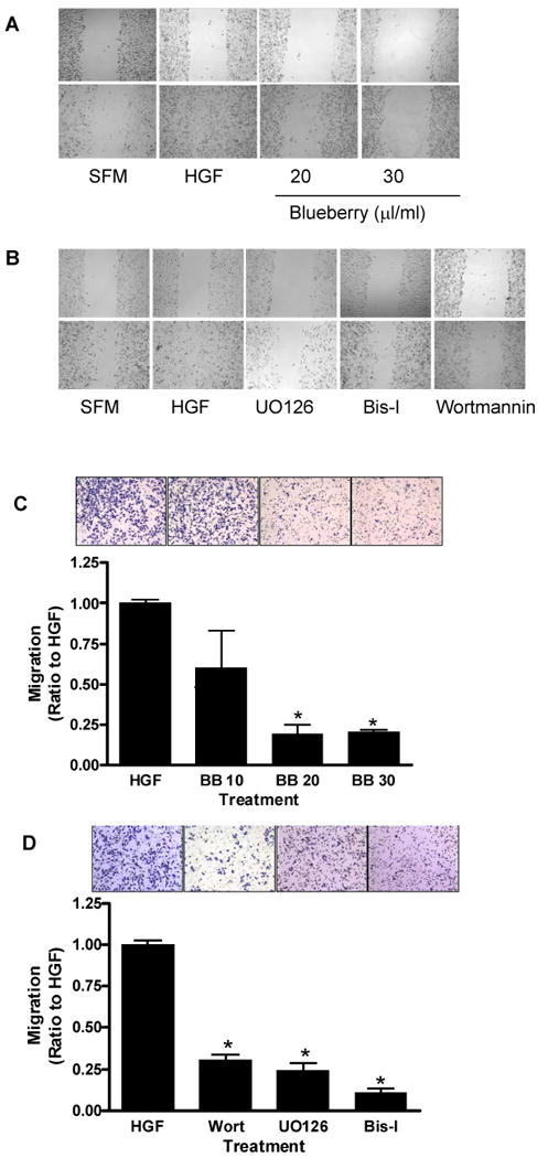
Confluent monolayers were scratched with a plastic pipette tip and incubated in serum free medium (SFM) in the presence of either HGF (40 ng/ml), or HGF plus blueberry (BB) extract (20 and 30 μl/ml) (A) or various cell signaling inhibitors (B) for 24 h. Migration of MDA-MB-231 cells through a PET membrane (0.8 micron Transwell culture inserts) was evaluated in the presence or absence of blueberry (C) or various cell signaling inhibitors (D) after 24 hours of treatment. Quantification of the number of migrating cells from three separate experiments. Data is expressed as a ratio to HGF-treated cells; mean ± SEM. Asterisk indicates significant difference from HGF-treated cells (n ≥ 3, p ≤ 0.01).