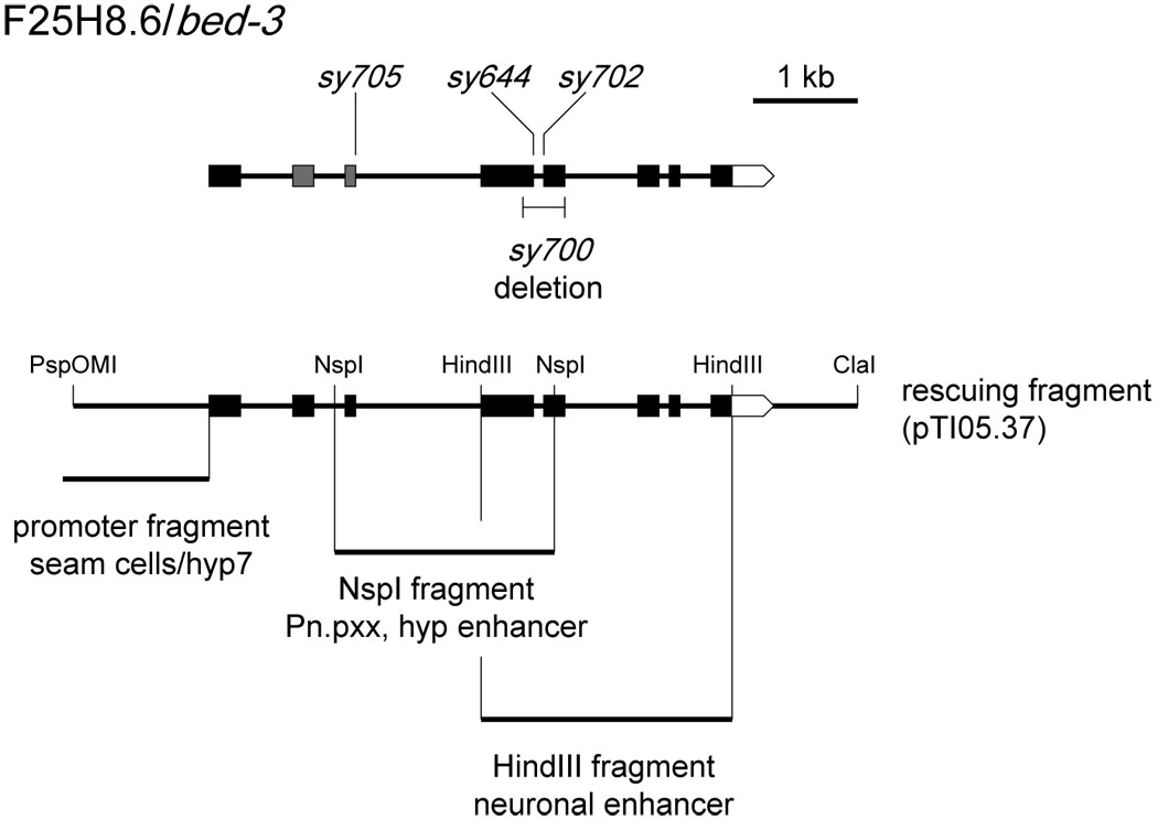
Figure 2. bed-3 gene structure.

Top: the intron/exon structure of bed-3 and locations of mutations. The BED Zn-finger domain is encoded in second and third exons (gray). Bottom: genomic regions tested for promoter and enhancer activity. The promoter fragment, placed upstream of a gfp reporter, drives expression in seam and hyp7 cells. When placed upstream of the promoter fragment, NspI and HindIII fragments drive additional expression, demonstrating the presence of enhancer activity.
