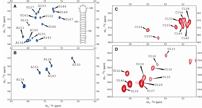
Fig. 1.
Natural abundance 1H-13C-HMQC spectra of the 35-nucleotide guanine riboswitch terminator hairpin. a C8H8 region of purine residues; the C8H8 signal of loop residue A138 is shifted just out of displayed spectral window (142.6 ppm, 8.264 ppm); the relatively high ppm value of H8 indicates de-stacking of this loop residue, which is also evident from the relatively high H2 ppm value seen in panel B. Also the sequence and secondary structure of the terminator hairpin is shown; numbering is according to Mandal et al. 2003 b C2H2 region of adenine residues c C5H5 region of cytosine residues d C5H5 region of uracil residues

