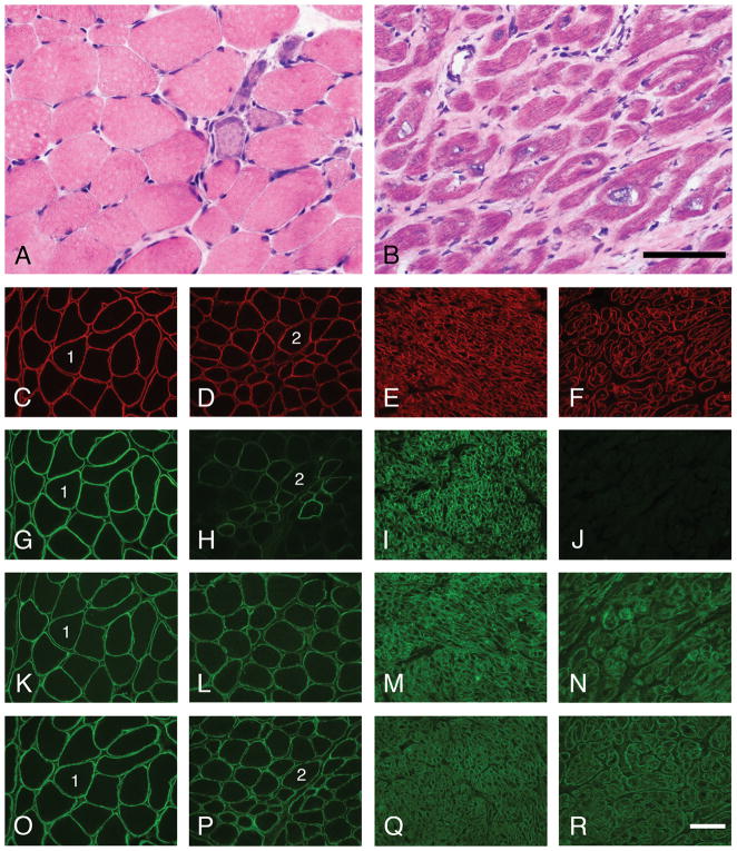
Figure 2.
Skeletal muscle and cardiac cryosections from patient #1 and control patients were stained with H&E and a panel of antibodies using immunofluorescence methodology. While the muscle pathology in patient #1 is mild for LGMD 2I (A), the heart shows features of cardiomyopathy (variation in myocyte diameter, variation in nuclear size and shape, and fibrosis) that are relatively severe (B). Staining for dystrophin, β-dystroglycan, and the core domain of α-dystroglycan is normal in both the skeletal muscle and the heart of patient #1. In contrast, staining for glycosylated α-dystroglycan ranges from negative to normal in a mosaic pattern in patient #1’s skeletal muscle and is absent in her heart. Shown here are representative micrographs of staining with H&E (A–B) and dystrophin (C–F), glycosylated α-dystroglycan (VIA4-1, G–H; IIH6, I–J), core α-dystroglycan (GT20ADG, K–N) and β-dystroglycan antibodies (O–R). Control skeletal muscle, panels C, G, K and O. Patient #1’s skeletal muscle, panels A, D, H, L and P. Control heart, panels E, I, M and Q. Patient #1’s heart, panels B, F, J, N and R. Scale bars, 100 μm.

