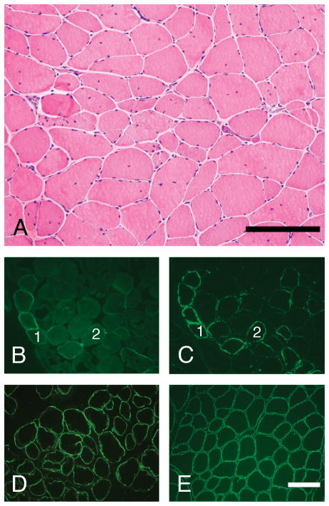
Figure 3.
Skeletal muscle cryosections from patient #2 were stained with H&E and a panel of antibodies using immunofluorescence methodology. The muscle pathology is relatively mild for LGMD-2I (A), but more severe than in patient #1 (compare with Fig. 2A). Staining for the core domain of α-dystroglycan (D) and β-dystroglycan (E) is normal. In contrast, staining for glycosylated α-dystroglycan ranges from negative to normal in a mosaic pattern that is characteristic for LGMD-2I (VIA4-1 antibody, panel B; IIH6 antibody, panel C). Scale bars, 200 μm.

