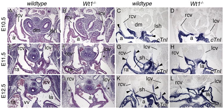
Figure 2.
Developmental onset of sinus horn defects in Wt1−/− hearts. Histological and molecular analyses of sinus horn development were carried out on transverse sections of the venous pole region of E10.5 to E12.5 wildtype and Wt1−/− hearts as indicated. Haematoxylin and eosin stainings (A,B,E,F,I,J), in situ hybridization analysis of cTnI expression on adjacent sections (C,D,G,H,K,L). Arrowheads point to myocardialized sinus horns. Asterisks mark subcoelomic mesenchyme in the mutant. a, common atrium; dm, dorsal mesocardium; la, left atrium; lcv, left common cardinal vein; lsh, left sinus horn; ra, right atrium; rcv, right common cardinal vein; rsh, right sinus horn; sh, sinus horn; vv, venous valves.

