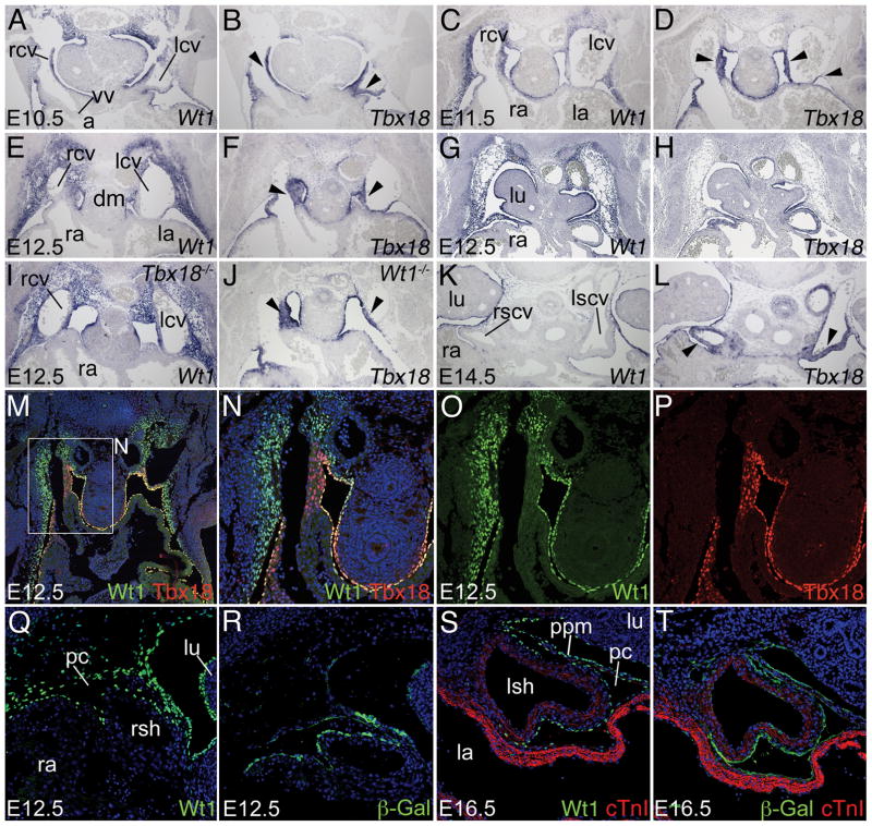
Figure 4.
Wt1 expression in the subcoelomic mesenchyme. A–L, Comparative in situ hybridization analysis of Wt1 and Tbx18 expression on transverse sections of the venous pole region of E10.5 to E14.5 hearts of wildtype (A–H,K,L), Tbx18−/− (I) and Wt1−/− (J) embryos. Black arrowheads point to Tbx18 expression in the mesenchyme and myocardium of the sinus horns. M–P, Immunofluorescence analysis of Wt1 and Tbx18 protein at the cardiac venous pole. Q–T, Comparative immunofluorescence analysis of Wt1 and β-galactosidase protein expression in the venous pole region in Wt1BAC-IRES-EGFPCre/+; Rosa26LacZ/+ embryos. cTnI immunofluorescence (red) marks sinus horn and atrial myocardium. Stages, genotypes and probes are as indicated. Abbreviations are as in Figures 1 and 2.

