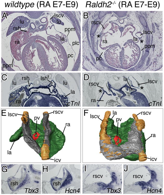
Figure 7.
RA-signaling is required for cardinal vein formation after E9.5 in the mouse heart. Histological, molecular and morphological analyses of sinus horns (A–F) and the SAN (G–J) were carried out on transverse sections of the venous pole region of E14.5 wildtype (left column) and Raldh2-deficient embryos food supplied with RA between E7 and E9 (RA E7–E9) (right column). A–F, Histological stainings with haematoxylin and eosin (A,B), in situ hybridization analysis of cTnI expression (C,D), and 3-D reconstructions of serial sections stained for cTnI in a dorsal-posterior view. Atrial myocardium is shown in green, cardinal vein myocardium in grey, the lumen of the cardinal veins in brown, and the pulmonary vein myocardium in red (E,F). Asterisks (B,D) mark persisting subcoelomic mesenchyme. G–J, in situ hybridization analysis of sections through the base of the cardinal veins for expression of SAN marker genes with probes as indicated. Abbreviations are as in Figure 1 and 2.

