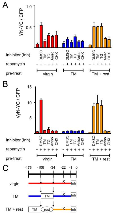
Figure 8. Effects of agents that induce acute and chronic protein folding stress on BiFC complex formation by FKBP and FRB fused to different fluorescent protein fragments.
Cells that expressed (A) FKBP-YN and FRB-YC or (B) FKBP-VyN and FRB-YC were analyzed either without pre-treatment (virgin), after 3 days of culture with 25 ng/ml tunicamycin (TM) or after 3 days of culture with tunicamycin followed by 3 days of culture without tunicamycin (TM + rest). An hour before rapamycin induction, the cells were treated with 1 μg/ml tunicamycin (TM), 50 ng/ml thapsigargin (TG), 25 μg/ml anisomycin (Aniso), or 50 μg/ml cycloheximide (CHX). The cells were harvested 3 hours after rapamycin induction and analyzed by flow cytometry. The bar graph shows the mean ratio of BiFC to CFP fluorescence. (C) Diagram depicting the experimental protocol used in parts A and B. The arrows indicate splits of the cells and the X indicates the time plasmids were transfected into the cells.

