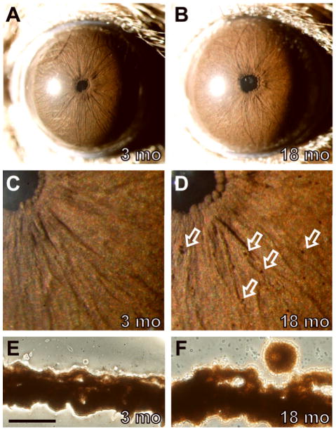
Figure 1.
Normal iris phenotypes of wild-type C57BL/6J mice. Comparisons of a representative 3-mo eye (left column) to an 18-mo eye (right column). (A) The normal C57BL/6J iris of young mice as viewed with broadbeam illumination originally imaged at 25X magnification. The iris is characterized by a sienna-brown color, a complex surface morphology with several small underlying vessels, and a circular pupil. The bright white circle to the left of the pupil is a reflection from flash photography. (B) With age, a number of clump cells are present on the surface of the iris and the iris becomes slightly more reddish in color. (C,D) At higher 40X magnification and less image reduction, clump cells are more readily visible, each casting a characteristic small crescent shadow (several are indicated by open white arrows, others in the field are unmarked). (E,F) Unstained cryosections of the same eyes shown above showing the presence of a melanin engulfed phagocytic clump cell in panel F on the surface of the iris stroma as a normal consequence of aging. These cells were not visible in 0/64 sections from the young eye in E and were present on the iris in 6/70 sections analyzed from the aged eye in F. Scale bar = 50 μm.

