Fig. 1.
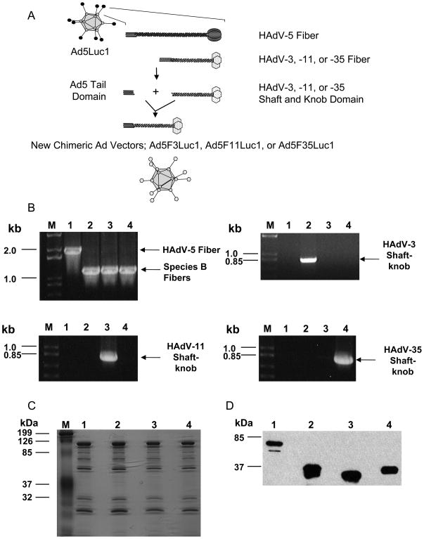
Characterization of chimeric Ad vectors. A: Schematic representation of a chimeric fiber of HAdV-3, HAdV-11, and HAdV-35 with HAdV-5 tail domain. B: Validation of the replacement of an HAdV-5 fiber gene with that of either HAdV-3, HAdV-11, or HAdV-35 in the HAdV-5 genome by PCR; all panels: M: DNA size markers; Lane 1: PCR using DNA from purified Ad5Luc1 as a template; Lane 2: PCR using DNA from purified Ad5F3Luc1 as a template; Lane 3: PCR using DNA from purified Ad5F11Luc1 as a template; Lane 4: PCR using DNA from purified Ad5F35Luc1 as a template. Upper left panel: PCR with pan fiber-specific primers; upper right panel: PCR with HAdV-3 fiber shaft knob-specific primers; lower left panel: PCR with HAdV-11 fiber shaft knob specific-primers; and lower right panel: PCR with HAdV-35 fiber shaft knob-specific primers. C: Analysis of protein composition of viral particles by GelCode blue staining of a 12% SDS-PAGE gel. A total of 1010 VP of purified Ad5Luc1 and chimeric Ad vectors was loaded per lane. M: protein molecular mass markers in kilodalton (kDa); Lane1: Ad5Luc1; Lane2: Ad5F3Luc1; Lane3: Ad5F11Luc1; Lane 4: Ad5F35Luc1. D: Detection of fiber proteins incorporated into purified viral particles by Western blot with an antibody against HAdV-5 fiber tail. A total of 5 × 10⁹ VP of the purified Ad vectors were run on a 12% SDS-PAGE; separated viral proteins were transferred to a PVDF membrane and probed by an antibody against the HAdV-5 fiber tail (4D2). Lane1: Ad5Luc1; Lane2: Ad5F3Luc1; Lane3: Ad5F11Luc1; Lane 4: Ad5F35Luc1; protein molecular mass markers (in kDa) are indicated on the left.

