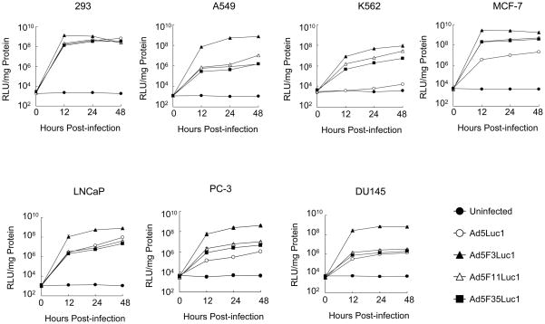
Fig. 6.
Comparison of transductional efficiency in various human cancer cells (HEK293, A549, K562, and MCF-7) and prostate cancer cells (LNCaP, DU145, and PC-3) with Ad5Luc1 and chimeric Ad vectors at various times. The luciferase activities in the lysates of cells infected with Ad5Luc1 (○) and chimeric Ad vectors Ad5F3Luc1 (▲), Ad5F11Luc1 (△), and Ad5F35Luc1 (■) at an MOI of 10 PFU/cell were measured at various time points (0, 12, 24, and 48 hours post-transduction) and normalized for protein concentration. Data points represent mean ± standard deviation (n = 3). Closed circles (●) indicate background luciferase activity.

