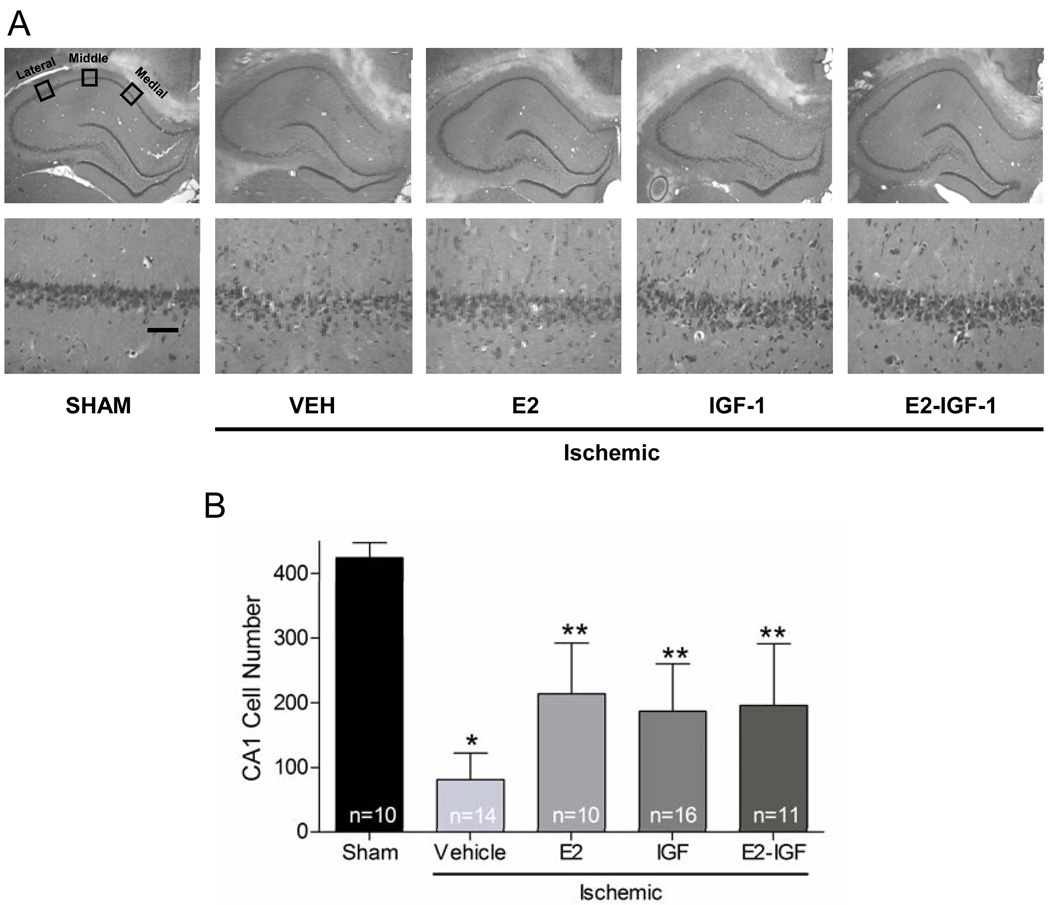
Figure 1.
Oestradiol and IGF1 provided significant protection against CA1 cell loss. (A) Representative photomicrographs from animals receiving sham surgery or ischemia treated with vehicle, oestradiol, IGF1, or oestradiol + IGF1 are shown at low (top, 4×) and high (bottom, 40×) magnification. Viable neurones were counted in 3 sectors (lateral, middle, and medial) in 4 haematoxylin and eosin stained sections of the dorsal hippocampus. Scale bar, higher magnification (40×) is 50 µm. (B) Data represent the grand sum (mean ± S.D.) of 3 counting sectors over both the right and left hemispheres. * Global ischemia induced significant CA1 cell loss in vehicle-treated compared to sham-operated animals (p<0.001). ** Treatment of ischemic animals with oestradiol, IGF1, or oestradiol + IGF1 provided significant neuroprotection compared to vehicle-treated ischemic animals (p<0.001).

