Fig. 4.
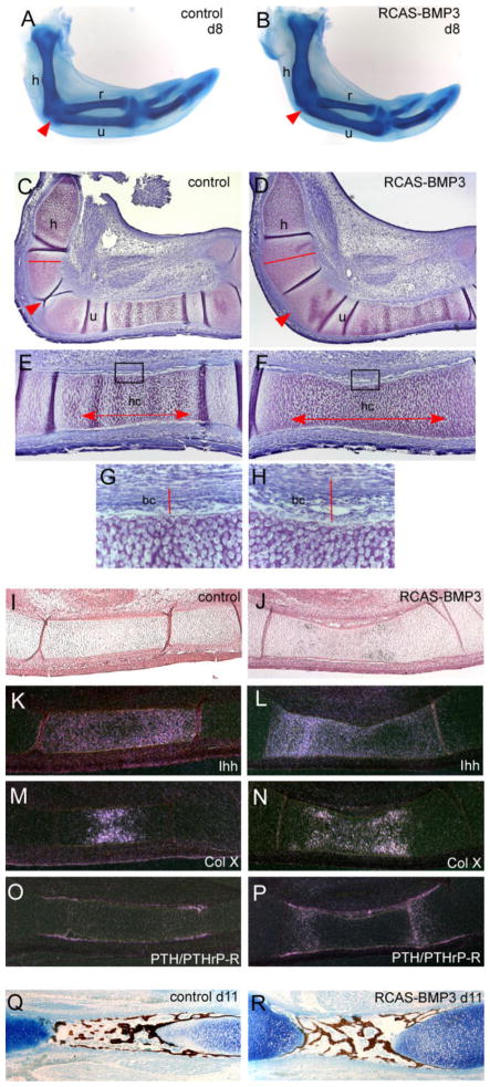
Effects of overexpressing replication competent avian retroviral vector–bone morphogenetic protein (RCAS-BMP3) in chick wings. A,B: Embryos were harvested after 8 days of development and the limb skeleton stained with Alcian blue. BMP3-infected wings have expanded cartilage elements and joint fusions at the junction of the humerus (h), radius (r), and ulna (u) (arrowhead). C,D: Toluidine blue stained sections through day 8 RCAS-BMP3–infected and contralateral control wings. Note the widening of the humerus (compare red line in D with C) and the lack of a joint (arrowhead) in the BMP3 injected cartilages. E,F: Higher-power view of BMP3-infected and contralateral control ulnas. The zone of hypertrophic chondrocytes is notable longer in BMP3 ulnas (compare arrows in F with E). The developing bone collar (black box) also appears wider. G,H: Higher power view of the boxed areas shown in E and F. The developing bone collar of the BMP3-infected ulna has an increased width when compared with the control (compare red line in H with G). I–P: Analysis of molecular markers in BMP3-infected ulnae and their corresponding contralateral controls on day 8 (HH 32) by radioactive section in situ hybridization. I,J: Bright field views of control and BMP3-infected ulna. K,L: Shows the expanded expression domain of Ihh in BMP3 ulna. M,N: Shows the expanded expression domain of collagen X in BMP3 ulna. O,P: Shows the increased expression of PTH/PTHrP receptor in chondrocytes and perichondrium/periosteum of BMP3 ulna. Q,R: von Kossa staining for mineral deposition in control and BMP3-infected ulna on day 11. Sections were counterstained with Alcian blue.

