
Figure 3.
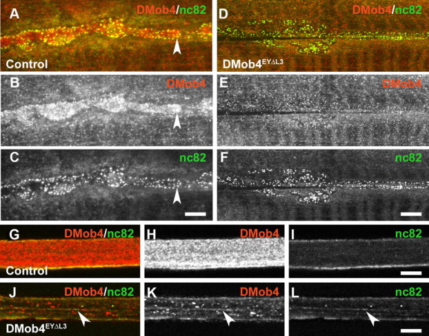
DMob4 expression in the peripheral nervous system of third-instar larvae. A–L, Confocal microscopy images of control (A–C, G–I) and DMob4EYΔL3 mutant (D–F, J–L) animals labeled with DMob4 antiserum (red) and counter-labeled with anti-bruchpilot (nc82) marker, which labels active zones at the NMJ and vesicular cargoes in peripheral nerves (green). Individual and merged confocal channels are presented as indicated. A–F, NMJs. DMob4 is present throughout synaptic boutons (B) and is enriched near active zones (concave arrowhead in A–C). DMob4 staining at the NMJ is absent in DMob4 mutants (E), indicating that NMJ immunoreactivity is specific to DMob4. Active zone staining persists in DMob4 mutants (F). G–L, Peripheral nerves. DMob4 is expressed at high levels in peripheral nerves and has both diffuse and punctate distribution characteristics (H). DMob4 staining of peripheral nerves is absent in DMob4 mutants (K), indicating that the DMob4 antiserum is specific. The distribution of nc82-positive vesicular cargo is altered in DMob4 mutants and not diffuse as observed in controls (arrowheads in J–L). Scale bars, 5 μm.