
Figure 5.
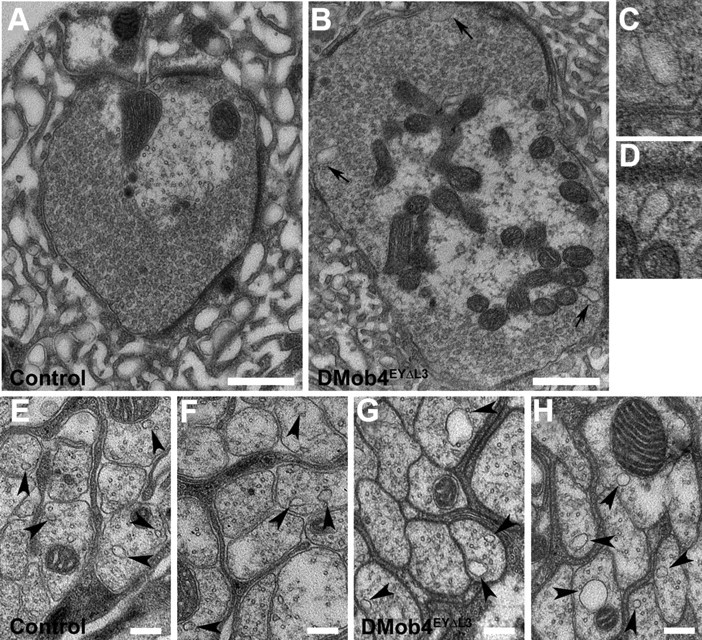
Transmission electron microscopy of DMob4 mutant NMJs and peripheral nerves. Control (A, E, F) and DMob4EYΔL3/Df(2R)42 mutant (B–D, G, H) NMJs. Large membrane-associated endocytic cisternae bud from DMob4 mutant NMJs (black arrows in B) but are absent in controls. C, D, High-magnification images of cisternae indicated in B. E–H, Cross-sectional profiles of axons from peripheral nerves of control (E, F) and DMob4EYΔL3/Df(2R)42 mutants (G, H). Microtubule-associated vesicles (black arrowheads) in control animals are smaller and less numerous than in DMob4EYΔL3/Df(2R)42 mutants. Scale bars: A, B, 500 nm; E–H, 200 nm.