
Figure 9.
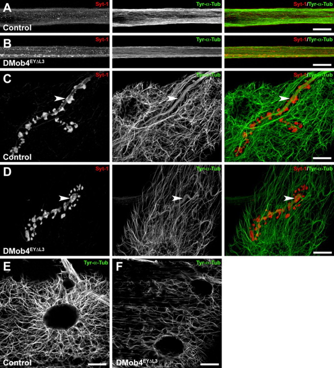
Diminished levels of tyrosinated microtubules in DMob4 mutants. A–E, Control and DMob4 mutants double stained for tyrosinated α-Tubulin (Tyr-α-Tub) using monoclonal antibody Tub-1A2 (green) and costained for Syt-1 (red). Individual and merged channels from confocal stacks are presented. A, B, Peripheral nerves. DMob4EYΔL3 homozygous mutants have decreased levels of Tyr-α-Tub, and individual microtubule bundles are less evident. In mutants, Tyr-α-Tub appears to have a smooth distribution. Syt-1 aggregates are evident in the DMob4 mutants (B). Stacks spanning half the diameter of a peripheral nerve are presented for increased resolution of microtubule networks. C, D, NMJs. Diminished levels of Tyr-α-Tub is observed in boutons of DMob4 mutant NMJs (compare arrowheads in green channel in C, D). Stacks of equivalent thickness spanning the NMJ are shown. E, F, Abdominal muscle. Microtubule network complexity, as well as Tyr-α-Tub levels are also reduced in the muscles of DMob4 mutants. Single confocal slices are presented. Scale bars, 20 μm.