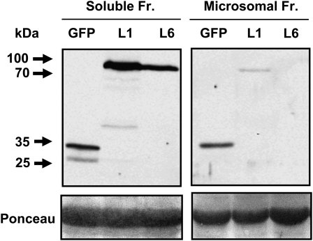
Figure 7.
Immunodetection of the AtPCS1 fusion protein. Abundance of AtPCS1:eGFP in the soluble versus microsome-enriched protein fraction (Fr.). Cell-free extracts of ΔPCS1 plants transformed with AtPCS1:eGFP, lines L1 and L6, and of a cytosolic GFP-expressing control line (GFP) were separated into the two protein fractions by differential centrifugation. Top, The protein samples (20 μg per lane) were analyzed by gel electrophoresis and by western-blot analysis using GFP-specific antibodies. Extracts of the GFP line served as a control. The arrows indicate the positions of protein standards, and their molecular masses are given in kD. Bottom, The Ponceau-stained membrane prior to immunodetection shows the large subunit of Rubisco as a prominent band and reveals contamination of the microsome-enriched fraction with soluble proteins.

