Fig. 2.
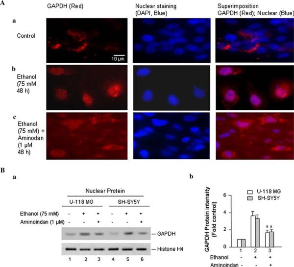
Effects of ethanol on the accumulation of GAPDH in the nucleus. Cells were treated by 75 mM ethanol for 48 h (for mRNA level and immunofluorescence) or 72 h (for protein level/Western blot). A Immunofluorescence microscopy was performed with anti-GAPDH antibody. U-118 MG cells were plated on a 4-well chamber slide and treated (a) without or (b) with 75 mM of ethanol or (c) with 75 mM of ethanol plus 1 μM of 1-R-aminoindan for 48 h. Then the cells were fixed and immunostained by mouse anti-GAPDH antibody and followed with fluorescein-conjugated anti-mouse secondary antibody (red). Stained slides were mounted in the presence of DAPI for nuclear staining (blue). The GAPDH (red) and nucleus (blue) and the merge of both GAPDH and the nucleus are indicated at the top. B Western blot analysis of nuclear GAPDH protein. (a) A representative of protein expression of GAPDH is shown. The anti-Histone H4 antibody was used as the loading control. (b) Quantitative analysis of Western blot results. A graph of the average optical density of GAPDH (normalized to the density of Histone H4) is shown. The relative intensity (relative optical density × pixel area) of autoradiographic bands from four independent preparations was evaluated using gel analysis software and a computer-assisted image analysis system. Data represent the mean ± SD of four independent experiments. *P < 0.001 compared with ethanol-treated group (without 1-R-aminoindan)

