
Figure 5.
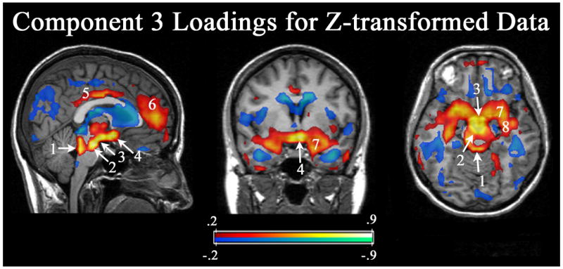
Principal Component #3. The 3rd component produced its highest loadings in the DA midbrain, the mammillary bodies and the hypothalamus. Note in the coronal and axial slices, a large area of the basal forebrain, including the extended amygdala also positively loads on this component. The axial slice passes through the mammillary bodies, which is slightly superior to the peak loadings in the substantia nigra/ventral tegmental area. Number labels: 1) colliculus/raphe, 2) substantia nigra/ventral tegmental area, 3) mammillary bodies, 4) hypothalamus, 5) posterior/mid cingulate, 6) medial frontal cortex, 7) amygdala, and 8) hippocampus.