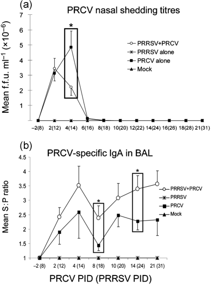
Fig. 6.
PRCV nasal shedding titres (a) and PRCV-specific IgA antibodies (b) in BAL of pigs co-infected with PRRSV and PRCV. (a) Nasal swabs were collected from each pig (n=39 at PRCV PID 2, n=31 at PID 4, n=20 at PID 6, n=15 at PID 8, n=11 at PID 10 and n=5–6 at PIDs 12–21 for PRRSV/PRCV-infected and PRCV only-infected pigs) at PRRSV PIDs 0–31 and tested by a cell culture immunofluorescence assay. The numbers of PRCV only-infected cells were expressed as f.f.u. ml−1. (b) BAL fluids were prepared at each PID from the numbers of pigs indicated in the legend of Fig. 2. PRCV-specific IgA antibody titres in BAL fluids were tested by ELISA. Each data point represents the S : P ratio (mean±sem). *P<0.05 (statistically significant difference between the dual-infected and PRCV only-infected pigs by the Kruskal–Wallis test).

