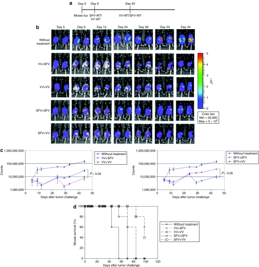
Figure 2.
In vivo luminescence imaging to demonstrate the antitumor effects generated by infection with SFV or VV in tumor-bearing mice. C57BL/6 mice were injected intraperitoneally with 5 × 10⁵/mouse of MOSEC-luciferase cells on day 0. Mice were then infected with 2 × 10⁶/mouse of SFV-WT or VV-WT on day 6, and the same dose of SFV-WT or VV-WT on day 25. The antitumor effects were characterized by luciferase expression using luminescence imaging. (a) Schematic diagram demonstrating the regimen of infection with SFV or VV. (b) Representative luminescence images depicting the antitumor effects in tumor-bearing mice infected with SFV/VV. (c) Line graphs depicting the fluorescence intensity in tumor-bearing mice infected with SFV/VV. (d) Kaplan–Meier survival analysis of MOSEC-luc tumor-bearing mice infected with SFV/VV. MOSEC, murine ovarian surface epithelial carcinoma; SFV, Semliki Forest Virus; VV, vaccinia virus; WT, wild type.

