Figure 3.
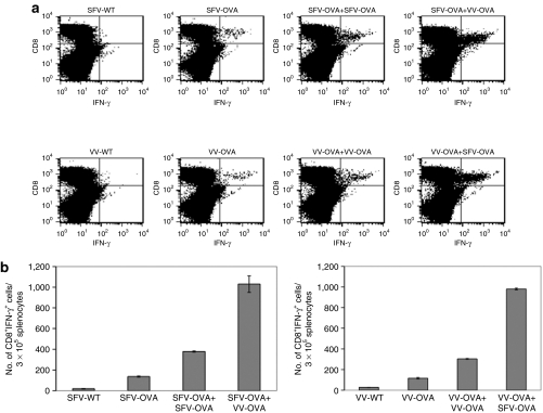
Infection with SFV-OVA followed by VV-OVA or vice versa leads to enhanced OVA-specific CD8+ T-cell immune responses. C57BL/6 mice were intraperitoneally injected with 5 × 10⁵/mouse of MOSEC cells on day 0. Mice were then infected with 2 × 10⁶ /mouse of SFV-OVA or VV-OVA on day 3, and the same dose of SFV-OVA or VV-OVA on day 17. The control group was injected with a single dose of SFV-WT, VV-WT, SFV-OVA, or VV-OVA on day 3. The spleens were harvested on day 24, the splenocytes were restimulated with OVA peptide SIINFEKL and the OVA-specific T-cell immune responses were characterized using intracellular cytokine staining followed by flow cytometry analysis. (a) Representative flow cytometry data depicting the number of OVA-specific CD8+ T-cells in the various groups. (b) Bar graph representing the number of OVA-specific CD8+ T-cells/3 × 105 splenocytes (mean ± SE). Data shown are representative of two experiments performed. IFN-γ, interferon-γ MOSEC, murine ovarian surface epithelial carcinoma; OVA, ovalbumin; SFV, Semliki Forest Virus; VV, vaccinia virus; WT, wild type.

