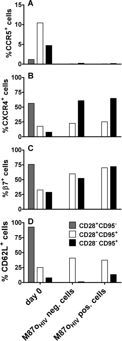
Figure 6.
Phenotype of M87oHIV-transduced CD4+ T cells. Peripheral blood from 4 or 5 naïve rhesus monkeys was used to determine the expression of CCR5 (A), CXCR4 (B), β7-integrin (C) and CD62L (D) on CD4+ T cell subsets prior to transduction and on day 7 after transduction and in vitro culture. The CD28 and CD95 paradigm was used to determine the lymphocyte maturation-associated T cell subsets. PBMC separated by Ficoll gradient were used to determine cell surface expression of β7-integrin, CXCR4 and CD62L on day 0. The cell surface expression of CCR5 on day 0 was determined by whole blood staining. The bars in all graphs represent median values.

