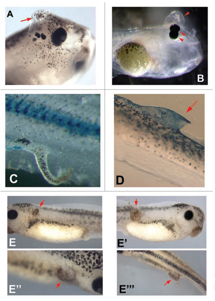
Figure 3.
Perturbation of growth in Xenopus embryos caused by manipulation of ion channels and electroporation. A variety of mRNAs encoding wild-type and mutant channels were microinjected into frog embryo blastomeres to screen for bioelectrical signals with roles in growth and pattern control. The VSOP167 proton channel (kindly provided by Yasushi Okamura) (A), the Cx32 gap junction subunit (plasmid kindly provided by Dan Goodenough) (B and C), and the HERG K+ channel (plasmid kindly provided by Annarosa Arcangeli) (D) result in ectopic growth and abnormal duplication of body structures such as eyes, sometimes forming extensive fin-like protrusions that are clearly associated with increases in cell growth. (E–E”’) Electroporation of embryos at stage 33, with no DNA, (95 msec interval, 5 msec pulse, 10X repeated) results in significant areas of ectopic growth 24 hours later. Red arrows indicate hyperproliferation.

