
Figure 4. Knockdown of UAP56 expression inhibits PE induced cardiomyocyte growth.
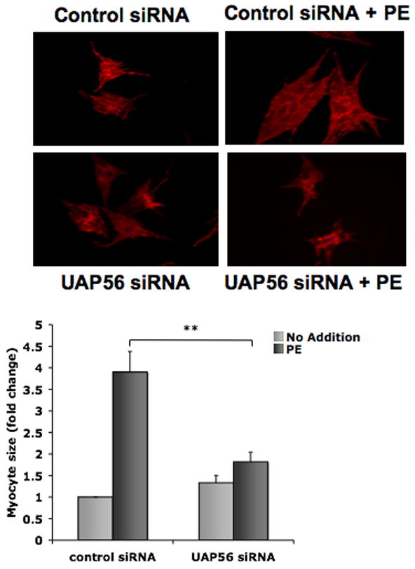
Cardiomyocytes were transfected with control or UAP56 siRNA for 72 hours and then treated with or without 50 μM PE for 4 hours. To assess cell size, cardiomyocytes were stained with a myocyte marker, sarcomeric α-actinin antibody, followed by Alexa 546 secondary antibody and fluorescence imaging was performed. Data are mean ± SEM. Cell area was measured for approximately 25 randomly chosen cells per condition. (**p<0.01).