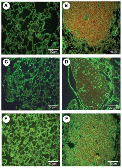
Figure 2.
The normal parenchyma in the lungs of the control animals shows a strong green birefringence of collagen V fibers (A). In contrast, the tumor parenchyma (B) shows a diffuse, green birefringence of collagen V fibers, which are organized in a reticular texture consisting of thin fibers that individually involve tumor cells and complex glands. The collagen III fibers show moderate green birefringence in the normal parenchyma (C) when compared to the tumor parenchyma (D). A strong green birefringence in the normal parenchyma (E) and tumor parenchyma (F) is shown by collagen I fibers. Immunofluorescence in A to F is at 400x magnification.

