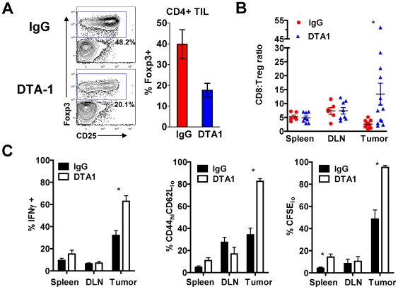
Figure 3. GITR ligation by DTA-1 limits Treg accumulation within the tumor and enhances intra-tumor CD8+ T-cell activity.
A. and B. B16-bearing mice treated with DTA-1 or IgG on day 4 had spleens, TDLN, and tumors harvested on day 10 and lymphocytes analyzed by FACS. A. Representative FACS plots with gate frequencies, (left) and mean +SEM for frequency of Tregs within live CD4+ TIL gate (right) are shown. B. Ratio of CD8+ to CD4+foxp3+ cells in spleen, TDLN, and tumor. *p = 0.05 compared with IgG tumor. Pooled data from 3 independent experiments are shown. C. Naïve C57BL/6 mice (n = 3−5/group) received 4×10⁶ CFSE-labeled pmel-1 Thy1.1+CD8+ T cells 1 day prior to B16 inoculation. Recipients received DTA-1 or IgG on day 4, and donor pmel-1 CD8+ cells analyzed in spleens, TDLN, and tumors on day 14. Mean frequency +SEM is shown for IFNγ+ (left), activated CD44hiCD62Llo phenotype (center) and proliferation by CFSE dilution (right) of transferred pmel-1 T cells is shown. For IFNγ recall assay (C, left), lymphocytes from spleen, TDLN, and tumor were re-stimulated for 6 hours with irradiated, gp10025-33 peptide-pulsed EL4 cells. Background IFNγ production for lymphocytes cultured with unpulsed EL4 cells was <1%. Over 85% of IFNγ+ cells were also CD107a+ (data not shown). * p<0.05 compared with IgG group. Representative of 3 independent experiments.

