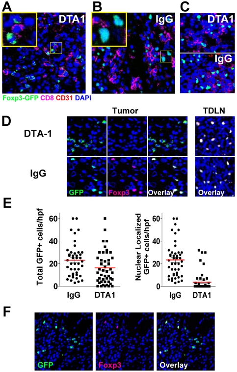
Figure 6. DTA-1-treated mice have abnormal intra-tumor Tregs which have lost foxp3 expression.
Tumors were harvested from day 10 B16-bearing foxp3 GFP mice (n = 4/group) treated with DTA-1 or IgG on day 4. Tumor sections were stained with anti-CD8 (magenta), anti-CD31 (red, to visualize endothelium), and DAPI (blue, for nuclear staining) and analyzed by immunofluorescence. A. DTA-1-treated tumor showing Tregs with irregular borders, weaker foxp3/GFP+ signal, and non-nuclear GFP localization (inset). B. IgG-treated tumor showing Tregs with foxp3 protein (GFP+, green) co-localizing with nucleus. C. DTA-1- and IgG-treated TDLN demonstrating intact foxp3+ cells. D. Left, top: DTA1-treated tumor co-stained with anti-foxp3. Note lack of foxp3 co-staining in cells with abnormal cytoplasmic GFP signal, compared with overlapping nuclear foxp3 and GFP in IgG-treated intra-tumor Tregs (left, bottom) or DTA-1- or IgG-treated TDLN (right). Scale for all images is show in A (bar = 25 µm). E. The number of GFP+ cells per high-powered field (hpf) were counted, regardless of either intensity or localization (“total”, left graph) or only with bright nuclear GFP signal (i.e. co-localizing with DAPI) (“nuclear GFP,” right graph). Pooled data from a total of 46 (IgG) or 48 (DTA-1) examined hpf (10–12 hpf per tumor ×4 tumors/group) are shown. F. Although the majority of DTA-1 treated Tregs have an “abnormal” GFP+ foxp3- phenotype, “intact” and “abnormal” Tregs can be found together within a minority of DTA-1 treated tumor sections.

