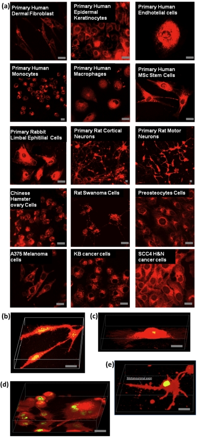
Figure 4. Selected examples of cells treated with Rhodamine-loaded polymersomes.
(a) Live primary human dermal fibroblast (HDF), primary human epidermal keratinocytes (HEK), primary human endothelial cells (HE), primary human monocytes (HMC), primary human macrophages (HMP), primary human mesenchymal stem cells (HMSc), primary rabbit limbal epithelial (RLE) cells, primary rat cortical neurons (RCN), primary rat motor neurons (RMN), Chinese hamster ovary (CHO) cells, rat Shawnoma (nemap22) cell, preosteocytes (MLO-A5) cells, human melanoma (A375SM) cell, human head & neck cancer (KB) and (SCC4) cells have all been exposed to Rhodamine-loaded polymersomes and successfully stained. 3D reconstruction from confocal laser scanning optical stacks of HDF cells (b), HE cells (c), SCC4 cells (c), and RMN cells (d). Figure bar = 0.02 mm.

